
From Clinic to Coffee Table: MyTechDailyNews Breaks Down the Science, Limits, and Safety of At-Home Fetal Dopplers
Some of the biggest changes in consumer technology are the quiet ones. A capability that once required a clinic appointment becomes a battery-powered wand with a small speaker, and it shows up in ordinary households. The at-home fetal doppler follows that pattern almost exactly: the rhythmic sound of a baby's heart, previously heard mainly during an OB-GYN visit on dedicated professional equipment, can now be produced on a couch at a moment the family picks.
Viewed only as a product story, that looks like a pure win for access. The more useful reading, in our view at MyTechDailyNews, is that this gadget is a diagnostic instrument re-engineered for consumers — and the gap between those two identities is exactly where confusion, false reassurance, and unnecessary panic collect.
Here is what the device actually captures, why gel is mandatory rather than cosmetic, how a mass-market unit differs from the tool a midwife carries, when sound usually becomes detectable, what the competing rhythms mean, what regulators advise, and where bonding stops and medicine begins.
Sound, Not Pictures: What the Handheld Probe Captures
Every fetal doppler rests on the Doppler Effect, the same phenomenon that makes an ambulance siren drop in pitch as it passes. The probe sends high-frequency sound waves through the abdomen. When those waves bounce off something in motion — a tiny heart, for example — they return at a slightly shifted frequency, and the electronics translate that shift into audio. What families hear is the familiar whoosh‑whoosh.
Consider what that description omits: there is no image. Clinical ultrasound produces detailed pictures of the fetus and the structures around it, while a doppler delivers sound only. The home device lets you listen rather than see, and that single design choice explains both its convenience and its limits.
Conductive gel is the most underestimated step. It is not lubrication for comfort. Air conducts high-frequency sound poorly and scatters waves before they reach tissue — a problem framed by acoustic impedance. Gel clears the air gaps between probe and skin, reduces impedance mismatches, and creates efficient coupling. Skip it and the returning signal scatters and weakens, making detection unreliable.
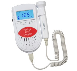 Figure 1: A pocket fetal doppler with a 3-MHz probe collects low-amplitude Doppler-shifted signals from the fetal heart. Source: KernMed
What $40 Buys Compared With What a Clinic Uses
Dopplers are not interchangeable. A unit ordered online for around $40 and the equipment in a doctor's or midwife's bag sit at opposite ends of the capability range. Professional devices carry higher sensitivity, so they can pull faint signals from a very small heart earlier and with far more reliability.
Clinical hardware also adds noise-filtering circuits that suppress interference from maternal blood flow and body movement, and it is checked and recalibrated on a regular schedule. Consumer units rarely see calibration at all.
At-home models are simplified, mass-produced derivatives. They can locate a heartbeat, but they are more exposed to false positives — mistaking the mother's own pulse for fetal activity, for instance — and to false negatives when the probe is mispositioned. They offer emotional reassurance; they do not offer clinical precision.
| Comparison | At-home doppler | Medical-grade doppler |
|---|---|---|
| Access | Purchased online for around $40 | In a doctor's office or a midwife's bag |
| Sensitivity | Simplified, mass-market hardware | Higher sensitivity for faint signals |
| Interference | More false positives and false negatives | Noise-filtering circuits suppress interference |
| Accuracy | Rarely or never calibrated | Calibrated on a regular schedule |
Timing and Anatomy: When a Heartbeat Usually Becomes Audible
For most households, dependable detection begins between weeks 12–16, the window in which these simplified units start catching the sound consistently. Detection at weeks 8–12 is possible in a clinician's office with medical-grade equipment, but it is often unreliable at home, so early success should not be assumed.
Anatomy explains the delay. During 8–11 weeks the uterus still sits deep inside the true bony pelvis, making acoustic detection difficult even with professional instruments. Around weeks 12–14 the uterus rises into the abdominal cavity; once it clears the pelvic brim, the fetal heart falls within practical reach of a transducer, and home pickups begin to work.
Even inside that sweet spot, variables interfere. An anterior placenta, attached along the front of the uterine wall, behaves like an acoustic muffler that dampens returning signals. Denser maternal tissue or a higher BMI lengthens the path the waves must travel and weakens them. A retroverted uterus tilts backward rather than forward, changing the probe's angle of approach and demanding more patience and precision. Fetal position and the early edge of gestation cause further misses — which is why silence should not automatically read as a warning.
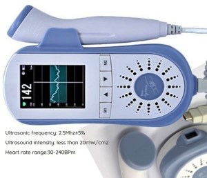 Figure 2: A compact fetal doppler monitors fetal heart rates during routine examinations for obstetricians, midwives, and expectant mothers. Source: pregnancy & baby.ie
Three Rhythms You Might Hear, and How to Tell Them Apart
The hardest part of home use is not physics but interpretation. Mixing up the mother's circulation with the baby's heart — or the reverse — produces either false reassurance or needless alarm, which is why clinicians and engineers alike stress learning distinct audio signatures.
- Fetal heartbeat: rapid, crisp and rhythmic, commonly compared to a galloping horse or a distant locomotive. Fetal cardiac activity typically runs between 110–160 beats per minute; recorded as a range, that is 110–160 beats per minute (BPM).
- Placental souffle: maternal blood rushing through the uterine arteries. It tracks the mother's pulse, usually 60–100 BPM, and arrives as a soft, low-pitched, synchronous swishing or swooshing.
- Umbilical cord flow: blood moving through the umbilical arteries. It matches the fetal rate but sounds sharper and mechanical — a whistling or whooshing instead of a clean cardiac thump.
Those overlaps are exactly why the field talks about filtering algorithms and signal processing: separating sounds that occupy the same acoustic space is a computation, not a listening trick. For users the lesson is humbler. A heartbeat is a signal buried in a complex acoustic environment, and a speaker on a couch does not replace training.
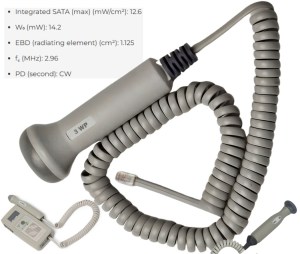 Figure 3: A waterproof 3-MHz fetal probe supports monitoring in early pregnancy. Source: Newman Medical
Under the Hood: The Engineering Trade-offs
Behind the familiar whoosh-whoosh is a tightly tuned hardware chain. Designers build an analog front end (AFE) around continuous-wave architectures working in the 2–3 MHz band. Most handheld probes use a dual-element piezoelectric transducer to isolate signal paths: on the transmit side, high-frequency driver circuits actuate the transmit crystal with high efficiency; on the receive side, a dedicated low-noise amplifier (LNA) pairs with an in-phase/quadrature (I/Q) demodulator to extract microvolt-level return voltages from abdominal tissue.
Once those reflections are demodulated into the baseband audio spectrum (< 3 kHz), a low-power microcontroller or DSP runs real-time autocorrelation to filter out maternal vascular artifacts and calculate fetal heart rate. Safety is the hard boundary: strict isolation barriers and capped driver thermal limits keep acoustic intensity below 20 mW/cm².
Frequency choice is a practical compromise. Most handheld units operate between 2–3 MHz. A 2-MHz probe penetrates deeper, which suits later gestational stages or patients with a higher BMI; 2 MHz buys reach, while 3 MHz buys clearer resolution at shallower depths, the reason early-pregnancy designs often prefer it. MHz selection, in other words, is a design decision rooted in anatomy and physics.
Architecture matters as well. Handheld dopplers mostly rely on continuous wave (CW) ultrasound: one crystal transmits continuously while another receives, producing a steady stream of reflections that converts cleanly into audio. Diagnostic imaging systems instead use pulsed wave (PW) ultrasound, alternating between sending and receiving. PW allows depth resolution and visual mapping, as in B‑mode scans, while CW stops at audio feedback with no imaging.
Why Regulators Push Back, and What ALARA Means
The FDA has long emphasized that fetal dopplers should be used only by, or under the supervision of, healthcare professionals. The worry is not mainly the waves themselves — ultrasound is generally safe at diagnostic levels — but misinterpretation and the delayed medical care that can follow. Prolonged or improper use can also theoretically cause slight tissue heating.
The guiding ethic for engineers is ALARA: As Low As Reasonably Achievable. Borrowed from radiation safety and applied to ultrasound, it shapes output power, duty cycles and probe design, and it supports short listening sessions rather than prolonged scanning. Every extra milliwatt of acoustic energy and every unnecessary minute of scanning is exposure that can and should be reduced. For clinicians ALARA is a usage guideline; for manufacturers it is a design imperative.
Benefits, Costs, and the Emotional Reality
With realistic expectations, the appeal is genuine: an immediate sense of connection between appointments, a moment that partners and siblings can share, and some relief for parents who have experienced pregnancy loss.
The costs are equally real. Hearing a heartbeat does not guarantee that everything is normal, and depending on that single sound can delay urgent medical care when fetal movement decreases. On the other side, failing to find a heartbeat — usually because of fetal position, placenta placement or simple user error — can trigger panic with no medical basis. These devices can support bonding; they should never replace professional guidance.
Common Misconceptions
- "If I can hear it, we're fine." Audio detection is not a diagnosis and says nothing about the rest of the pregnancy.
- "Silence means something is wrong." Fetal position, an anterior placenta, or being at the early edge of gestation explain many empty sessions.
- "It's sold online, so it's a toy." It is a consumer version of diagnostic ultrasound, not a novelty gadget.
- "Gel is optional." Without it, air gaps scatter the waves and detection collapses.
- "Earlier detection is better detection." Home units become consistent later; the clinic, not the couch, is where earlier attempts work.
- "Scanning longer will find the answer." ALARA points to short sessions, not more exposure.
Checklist: A Shorter, Safer Listening Session
- Start with a full bladder, which helps lift the uterus.
- Lie flat before searching.
- Apply enough gel to remove air gaps.
- Begin low, near the pubic bone.
- Rock the probe slowly instead of sliding it quickly across the skin.
- Learn the slower thump of your own pulse and the rhythm of placental flow so you can tell them apart from a fetal beat.
- Expect the sound to be fast — usually 110–160 beats per minute — like a galloping horse or a rapid train.
- Keep the session brief and stop after a few attempts, in line with ALARA.
- Do not substitute the device for care: contact your provider instead of scanning repeatedly.
Frequently Asked Questions
How early can a handheld unit find a heartbeat at home?
Most families first hear it between weeks 12–16. Clinics using medical-grade equipment may detect around weeks 8–12, but the same attempt at home is frequently unreliable.
Can the device tell me whether the baby is healthy?
No. It captures sound instead of images, so it cannot display anatomy or confirm normal development. That is the line between listening and diagnosing.
Why does the heartbeat sound so fast?
Fetal cardiac activity normally sits at 110–160 beats per minute (BPM), which is why the audio reads as a gallop rather than a relaxed adult thump.
What is the slower swishing I sometimes pick up?
Usually the placental souffle — maternal blood moving through the uterine arteries, matching the mother's pulse at 60–100 BPM. Cord flow, by contrast, follows the fetal rate but adds a sharper whistling quality.
Are the sound waves themselves risky?
At diagnostic levels ultrasound is considered generally safe, but prolonged or improper use can theoretically cause slight tissue heating. Hardware caps intensity below 20 mW/cm², and ALARA keeps sessions short.
Should I buy one anyway?
If it is treated as a bonding tool, the experience can be meaningful. If it would be used to check on a baby after reduced movement, that is where the risks concentrate.
Action Advice: Use It as a Moment, Not a Monitor
- Treat any session as a bonding ritual with a time limit, not as a home health check.
- Set expectations around the calendar: the realistic window for consistent home listening opens later than most marketing suggests.
- Log what you hear — rate, rhythm, or silence — and bring it to your next appointment rather than interpreting it alone.
- Escalate to a healthcare professional for anything that worries you, instead of reaching for the probe.
- If you already own one, review the guidance with your provider so the device sits inside care rather than in place of it.
Seen from a general-tech angle, the handheld doppler is a compact case study in trade-offs: CW simplicity against PW precision, consumer accessibility against clinical calibration, reassurance against responsibility. The sound is real physics in practice — MHz-range reflections turned into an audible sign of life — but it is a signal shaped by uterine position, placentas, anatomy and regulatory guardrails.
MyTechDailyNews will keep covering this wider pattern, in which specialized instruments leave the clinic and land in everyday hands, and will keep reporting the same two things each time: what the gadget genuinely does, and the point where its convenience runs out.