
MyTechDailyNews General: AI Circled a 1.5 cm Spot on an Ordinary CT Scan — What That Means for Your Next Check-up
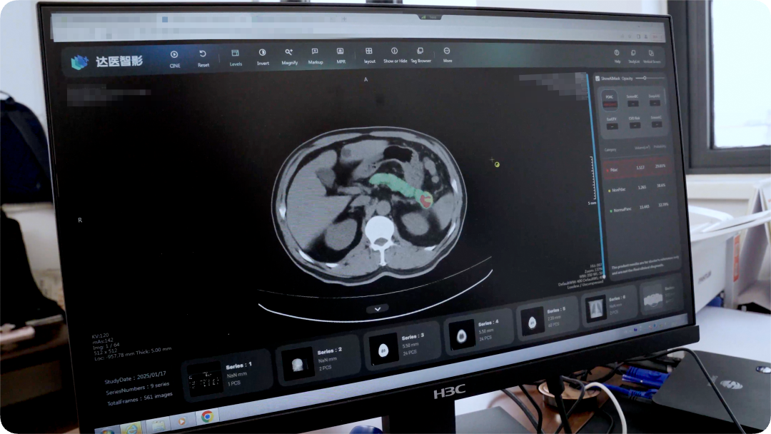
Some of the most consequential AI in healthcare right now is not doing anything new. It is simply re-reading work that has already been done. That is exactly what happened in a case from the Affiliated People's Hospital of Ningbo University, where a software model looked at a plain, no-contrast CT scan — imaging taken because a patient had complained of dizziness for three days — and marked a lesion roughly 1.5 cm wide in the pancreas, adding a note that it could be malignant. No one in the clinic had been looking for a pancreatic problem at all. On the MyTechDailyNews General desk we break down how that flag happened, why the same idea is now being stretched to several cancers at once, and why a second line of research, one that estimates survival odds from an ordinary face photo, is making even its own authors uneasy.
A complaint that pointed somewhere else entirely
Zhu Kelei, vice president of the hospital and a chief physician in hepatobiliary, pancreatic and general surgery, later described the moment of discovery: “This is something none of us doctors would have imagined, because the patient's chief complaint was dizziness, while pancreatic trouble usually shows up as abdominal pain.”
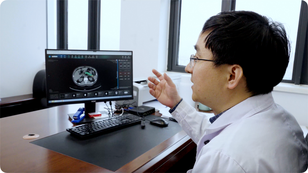
The alert redirected the whole visit. A contrast-enhanced CT was arranged immediately, and its result pointed clearly to pancreatic cancer. Mr. Qiu went into surgery soon afterward, and the pathology report confirmed a pancreatic ductal adenocarcinoma. His daughter put the outcome plainly: “We are so grateful — my father really got his life back.”
Pancreatic cancer carries its grim nickname for reasons this case makes visible. The gland sits deep inside the abdominal cavity, boxed in by the stomach, the duodenum and the spleen, and early disease produces almost no distinctive symptoms. By the time a patient feels unmistakably unwell, the cancer has frequently already spread in silence.
The scale of the problem explains the appetite for a shortcut. In China, more than 130,000 people are newly diagnosed every year; over 80% of them are already at an advanced stage at diagnosis, and the five-year survival rate remains below 10%.
Zhu's own summary of what the software added was understated: AI gave the doctors an unusual pair of eyes, one that examines the same patient from a completely different angle.
Opportunistic screening: reading the scans a hospital already has
The programme started in November 2024, when the Ningbo hospital introduced medical AI from Alibaba DAMO Academy, developed with the Lakeside Laboratory, and launched a prospective clinical trial together with Professor Jin Gang's team at the Shanghai Pancreatic Disease Institute — becoming the first hospital in Zhejiang Province to open a dedicated early pancreatic cancer screening clinic.
To date, the Ningbo effort has gone through CT imaging from more than 40,000 people and surfaced 8 pancreatic cancers, 6 of them at an early stage. All of those patients received surgery.
The method behind the PANDA model is the part worth understanding, because it explains why no extra test was required. The research team registered contrast-enhanced CT images from over 2,000 pancreatic cancer patients back onto plain CT, teaching the model to recognise subtle pancreatic lesions in lower-resolution pictures — precisely the images produced by the cheapest, most routine scan.
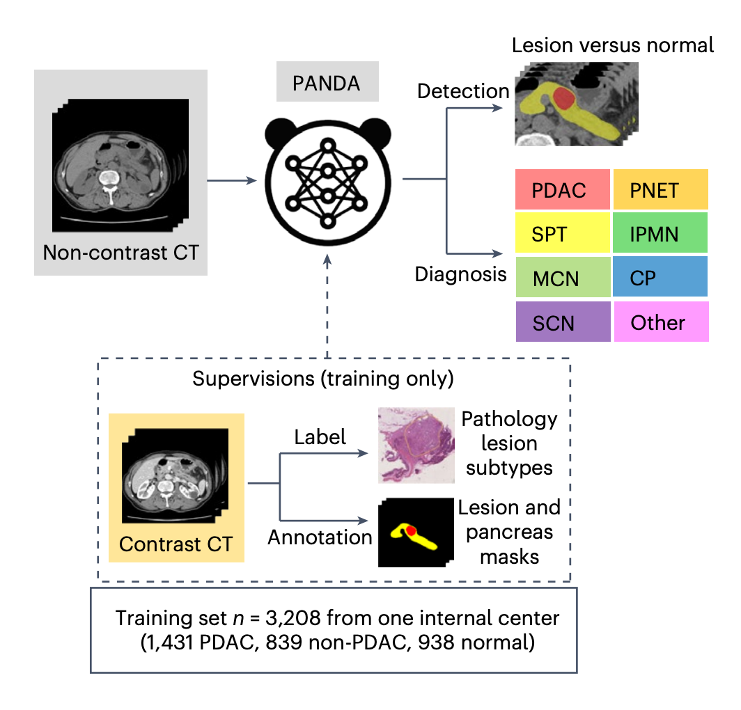
On those lower-resolution images the model identified pancreatic cancer patients with a sensitivity of 92.9% and a specificity of 99.9% — about 34% higher sensitivity than experienced radiologists reading the same kind of scan.
| What readers compare | Plain (non-contrast) CT | Contrast-enhanced CT |
|---|---|---|
| Contrast injection | Not needed | Required |
| Relative cost | Lower | Higher |
| Suitability for mass screening | Feasible — it is the most common imaging exam in outpatient clinics | Poor fit for large populations |
| Image resolution for the AI | Lower; PANDA was trained to work here | Higher; used to build the training pairs |
| Extra burden on the patient | None, if the scan already exists — this is what “opportunistic screening” buys | A booked, additional examination |
Because the approach rides on imaging that has already been captured, patients take on no additional examination burden: any ordinary clinic visit or routine physical check-up can become the window in which an early cancer is noticed.
The experiment is no longer confined to one hospital. DAMO Academy's multi-cancer early screening AI has begun global rollout, now covering 9 countries and regions and serving over 20 million person-times. The stated target is a “one scan, many screens” workflow: from a single plain CT, the AI would not only look for pancreatic cancer but also screen for liver, esophageal, stomach and colorectal cancers, plus acute conditions such as aortic syndromes.
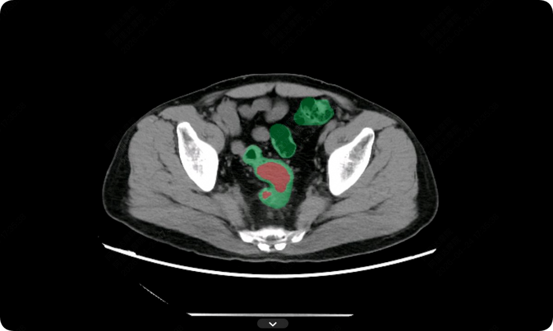
The gut is already the second published test case. After the pancreatic work, DAMO Academy released the colorectal screening model DAMO COCA, which went through plain CT images from 27,000 people and precisely picked out 5 colorectal cancers that had been missed, with 86.6% sensitivity and 99.8% specificity.
Why the machine noticed what a busy human did not
- Experience-based judgment and clinical decision-making remain irreplaceable in human doctors.
- But human eyes carry fatigue blind spots, and a stack of several hundred detail-heavy image slices is where omissions happen.
- Human attention is drawn to the complaint a patient walked in with — which is why a pancreatic region can stay unread when the story on the chart is dizziness.
- The software has no fatigue, is not pulled toward the chief complaint, and has no “I have read too many films today” state to recover from.
That difference is what surfaced a lesion that might otherwise have waited until late stage to announce itself.
The other experiment: reading survival odds from a photograph
AI's exploration of cancer does not stop at imaging. Recently, a research team at Harvard-affiliated Massachusetts General Hospital published a study in Lancet Digital Health in which one ordinary photo was used to predict a cancer patient's risk of death. The predictions were not fully accurate — but the patients the model scored as higher risk did in fact die at higher rates.
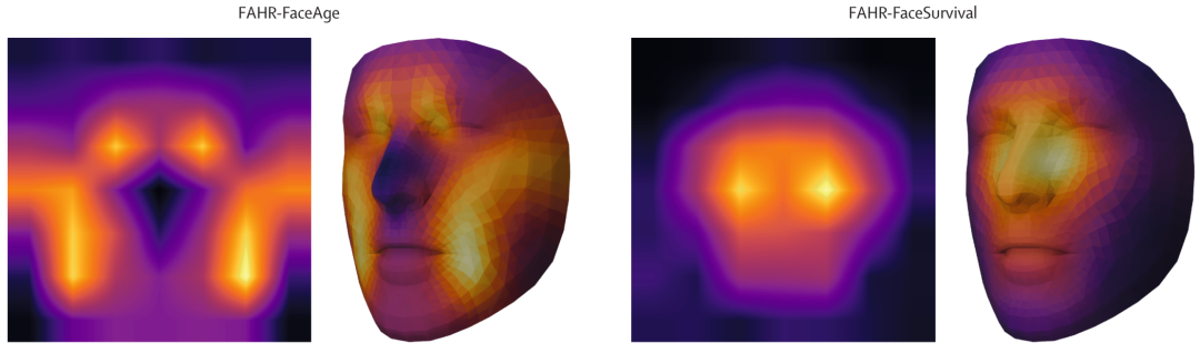
The logic rests on “biological age”. Two people who are both 60 years old may look 52 and 71, because lifestyle, disease history and genetics pull them apart. Researcher Mak notes that smokers tend to look older than their actual age, and heavy drinkers do too.
The training route is unusual. The team first enabled an algorithm to analyse and process 40 million ordinary face images without assigning it any specific task, then tuned that model to read faces in two different ways: one mode estimates a person's physiological age, the other predicts a cancer patient's odds of survival.
Where the model looks also splits. For physiological age it leans on regions such as the nasolabial fold — the line many of us call the upper cheek area — and parts of the forehead. For death-risk prediction it focuses more on the bridge of the nose and the area beneath the eyes.
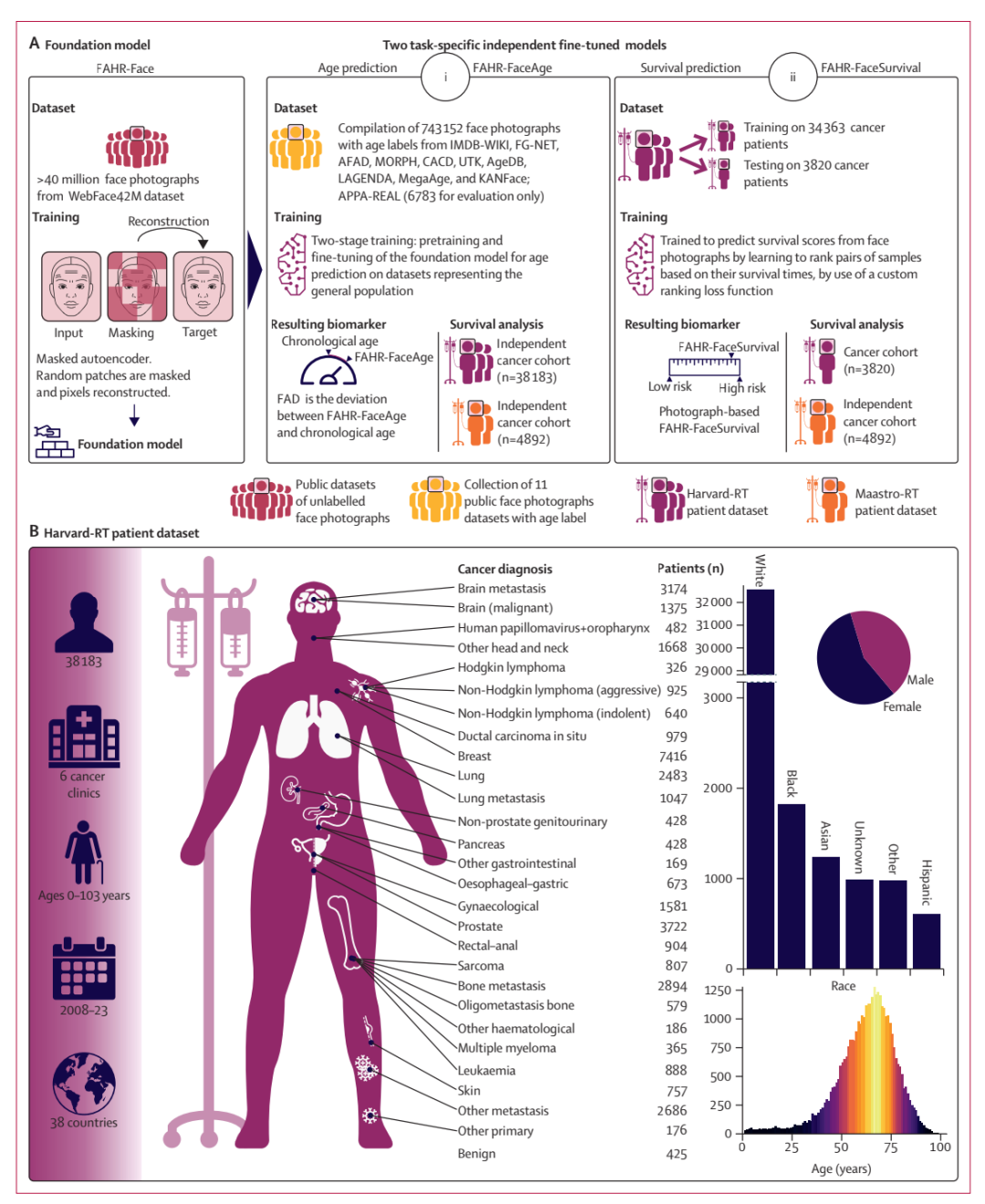
Surface variables seemed not to matter: whether the person was frowning or smiling, whether makeup was worn, and what the lighting was like did not shift the age estimate.
Common cosmetic surgery was also taken into account — with one quirk: people who have had a nose job tend to look slightly older in photos taken after the procedure than in the ones taken before it.
The numbers behind the risk score
- In a test group of 3,820 cancer patients, more than half of those in the highest death-risk group had died within 90 days of the photo being taken.
- In genetic data from lung cancer patients, FaceAge scores ran negatively correlated with CDK6, a gene that plays an important role in cell-cycle regulation and is involved in the progression of multiple cancers.
- The uncomfortable implication: a model trained only on faces appears to have captured, at some level, a signal that lives in the genome.
Separately, the chief executive of Sophont shared another case on social media, this one involving osteosarcoma. The company's model, Holon-S1, analysed the patient's pathology slide. It had never been trained specifically for osteosarcoma, yet it extracted a key piece of biological information: the patient's fibroblast-related genes were overexpressed.
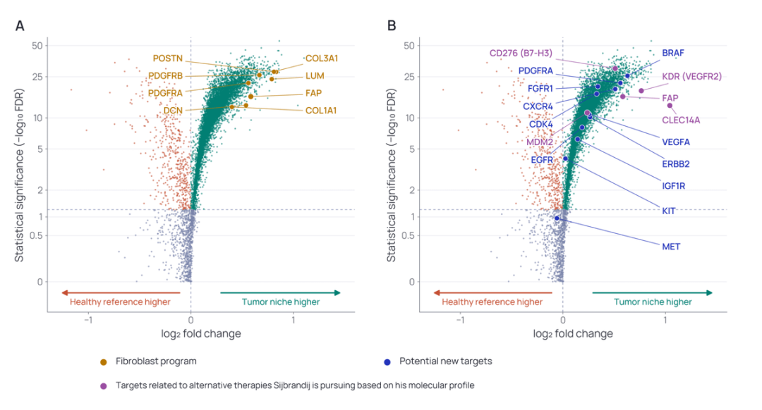
That same phenomenon had already been identified by the patient's own medical team through advanced molecular testing, which had also pinned down a matching treatment target.
The fairness problem hiding behind a friendly tool
FaceAge's researchers reserved space in their paper for a worry they could not shake: the tool may be a genuine help to clinicians, but it could also be extremely attractive to life insurers or employers looking to size up an individual's risk.
Watched side by side, the two cases are the same technology pointing in opposite directions. The moment the system pulled Mr. Qiu out of a crowd of patients who had come in for unrelated reasons was the benevolent version. A future insurance applicant tagged as high risk because of a single photograph is the version that opens a new set of problems.
There is a second, quieter risk. The “visual features of ageing” a model learns may not transfer across all skin tones and facial structures. If a model systematically overestimates biological age for certain ethnic groups, that error copies and magnifies inequality that already exists — in a way so hidden that the person misjudged never learns what happened.
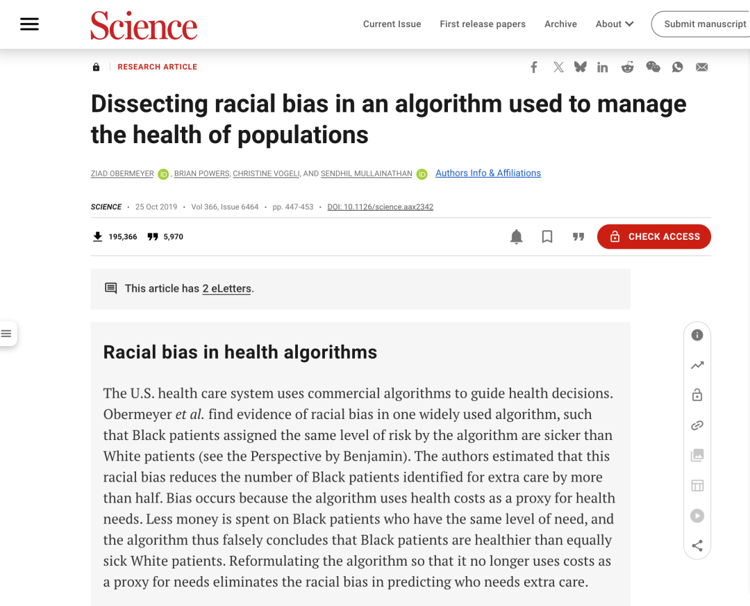
History supports the concern. Earlier research found that commercial models used to predict patients' medical needs systematically underestimated the needs of Black patients, because the models treated historical medical spending as a stand-in for health status — and Black patients with the same illness had historically been given fewer medical resources.
The researchers' own condition is explicit: before FaceAge reaches clinical use, it needs testing in far more diverse populations so that potential errors can be identified and removed.
Handling those questions, the study suggests, is considerably harder than training the model in the first place.
Common misconceptions readers run into
- “AI cancer screening means booking another test.” The point of opportunistic screening is the opposite: the AI reads a plain CT that already exists, so no extra examination is added to the visit.
- “If my complaint is unrelated, nothing else will be checked.” Mr. Qiu's record was dizziness for three days, and the flagged lesion sat in his pancreas — a site that typically presents as abdominal pain, not a head symptom.
- “92.9% sensitivity means the machine has replaced the radiologist.” The reported comparison is that the model reaches about 34% higher sensitivity than experienced radiologists on this task; experience-based judgment and clinical decision-making still belong to doctors.
- “A face-photo score must be fooled by makeup, expressions or bad lighting.” For FaceAge, frowning or smiling, wearing makeup or not, and the lighting environment did not change the age prediction; the model even accounts for common cosmetic surgery.
- “One trained model works equally well for everyone.” Learned ageing cues may not fit every skin tone or facial structure, and bias can compound existing disparities unnoticed.
FAQ: the questions that follow this story
Did the AI diagnose the cancer?
No. The system flagged a roughly 1.5 cm lesion and suggested a malignant possibility. Direction came next from a contrast-enhanced CT, and the final answer — pancreatic ductal adenocarcinoma — came from the pathology report after surgery.
Why could a scan ordered for dizziness turn up a pancreatic finding?
Because the plain CT the hospital already held was readable in that region. The model examined it from an angle no clinician was aiming at, which is precisely the gap “opportunistic screening” is designed to exploit.
Is this kind of screening available broadly?
The reported footprint is expanding: the Ningbo hospital has run its prospective trial since November 2024 as the first early pancreatic cancer screening clinic in Zhejiang Province, and DAMO Academy's multi-cancer early screening AI now covers 9 countries and regions with over 20 million person-times served. Whether your own provider offers it is a separate question to ask.
What is “one scan, many screens”?
It is the stated goal of reading a single plain CT for multiple targets at once — pancreatic, liver, esophageal, stomach and colorectal cancers, plus acute aortic syndromes. The colorectal model DAMO COCA is the published next step, with 86.6% sensitivity and 99.8% specificity after finding 5 missed cancers in 27,000 people's images.
Can a photo of my face be used against me?
That is the risk the FaceAge authors themselves raised: clinicians may benefit, while life insurers or employers may want the same score for risk assessment. The researchers' requirement before clinical use is validation in more diverse populations, with potential errors identified and eliminated.
Could the model be wrong specifically because of how I look?
Possibly. Its learned “ageing” features may not apply evenly to all skin tones and facial structures, and a systematic overestimate for some groups would replicate existing inequality invisibly. Precedent exists in commercial need-prediction models that under-served Black patients by using historical spending as a proxy for health.
Action suggestions: what to take from this
- Keep your own imaging records. Opportunistic screening only works when a plain CT already exists for a system to re-read, so having prior scans on hand matters.
- Ask directly whether an early pancreatic cancer screening clinic, or opportunistic AI re-reading of existing CTs, is available where you are treated.
- Do not wait for a symptom that matches your assumption about the organ. Pancreatic problems usually register as abdominal pain, and early disease is largely symptom-free.
- Treat an AI flag as a starting point, not a verdict: confirm through contrast-enhanced imaging and, where relevant, pathology, as the Ningbo team did.
- Cost context: contrast-enhanced CT involves an injection and higher expense and is unsuited to mass screening, while plain CT is the most common outpatient imaging exam — the reason it is the target of these models.
- If a photo-based risk tool appears in any non-clinical setting, ask two things before consenting: which populations it was tested on, and who is allowed to see the score.
Mr. Qiu's three days of dizziness ended in a 1.5 cm tumour caught early, and that is the version of this story everyone wants. The reporting behind it — a model trained to see through lower-resolution images, a second model trained to read risk through a face — also sketches what comes next: screening that costs you nothing extra, and measurement that may know things about you that you never submitted. Accuracy numbers are the easy half of that future. The harder half, as the FaceAge team says, is deciding what these scores are for, whose faces they were really tested on, and who gets to ask.
Written by Sarah Chen, Tech Daily. Tips, corrections and story ideas for the MyTechDailyNews General desk: hello@techdaily.com.